Do you want a profession that ultimately allows you the work/life balance, a haven to actually
enjoy your job and making it a meaningful matter other than just a paycheck?
Without mincing words, Radiography is the umbrella term for a truckload of career
opportunities. You’ve got various modalities to lay your eggs, perch on and an array of
professional branches to make a choice from.
These are the three basic specialties in Radiography and we’ll be taking a careful plunge,
explaining them dutifully:
1. Diagnostic Radiology (Medical Imaging)
2. Interventional Radiology
3. Radiation Oncology (Radiation Therapy)
To perfectly understand the differences between Radiography and Radiology, check:
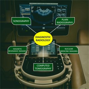
DIAGNOSTIC RADIOLOGY
Diagnostic radiology is a medical specialisation that involves undertaking a range of imaging
procedures to obtain images of the inside of the body. The diagnostic radiologist then
carefully interprets these images to diagnose illness and injury. This is where the various
radiographic modalities fall under.
Diagnostic imaging tests can include:
● X-rays (plain radiography)
● CT (computed tomography) scans
● MRI (magnetic resonance imaging) scans
● Ultrasound examinations (also known as sonography)
● Nuclear medicine imaging techniques.
X-rays
X-rays are a form of electromagnetic radiation (a.k.a. photons; light particles) that were
discovered in 1895, giving birth to the field of radiology. X-rays consist of ionizing radiation
generated from an x-ray machine that pass through patients and are used to create images
of whatever they pass though (people, carry-on bags, teeth, animals, etc.). Images are
created by the x-rays that penetrate travel through the object being imaged and reach a
detector on the other side. The images are referred to as “radiographs” or “plain films.”
Fluoroscopy
If an x-ray machine is like a camera, then fluoroscopy is like a video camera – x-rays are
produced in a pulsed or continuous fashion and generate real-time images of the body.
Think of it as “video x-ray.” The images are of much lower quality than conventional
radiographs to limit the patient’s overall radiation exposure.
Fluoroscopy allows radiologists and physician extenders to see what’s happening real time.
It is often used to guide procedures, such as lumbar punctures and injections and determine
when to take a true radiograph when evaluating the gastrointestinal (GI) and genitourinary
(GU) tracts.
Computed Tomography (CT) Scan
Computed tomography (CT scan) is a form of imaging that contains a donut-shaped ring
(gantry) of x-ray generators that rotates around the patient, getting information from multiple
different angles. This produces a stack of two-dimensional (2D) cross-sectional images of
the body that can be combined to create three-dimensional (3D) appearance. CT scans
rapidly obtain high quality images of the body and are capable of scanning the entire body in
under a minute. Because of this, CT is a workhorse of medical imaging, particularly in the
emergency setting.
Ultrasound
Ultrasound is a non-ionizing form of radiology that uses sound waves to create images of the
inside of the body. The sound waves are created by the ultrasound probe (aka transducer),
which enter the body, interact with various tissues, and return to the probe where the sound
waves are detected. A computer converts those sound waves into images.
Ultrasound is often used to visualize organs such as the liver, gallbladder, kidneys, spleen,
portions of the pancreas, uterus, ovaries, etc. Ultrasound can also be used to visualize blood
vessels (flow direction, speed, and even waveform), evaluate hernias and joints, and to
assess the health of fetuses (unborn babies).
Magnetic Resonance Imaging (MRI)
Magnetic resonance imaging (MRI) is a non-ionizing form of radiology that uses magnetic
fields and radio waves to create images of the inside of the body. It’s like ultrasound’s cooler
cousin. Of all imaging modalities, MRI is by far one of the most fascinating.
MRI uses gadolinium-based contrast agents similar to CT’s iodinated contrast. This makes
pathology stand out and easier to diagnose.
Nuclear Medicine
Nuclear medicine is occasionally referred to as “unclear medicine”. It involves giving patients
a radioactive medication via IV or by mouth. The radioactive drug will go through a
physiological pathway in the body and “trace” that pathway. Hence, radioactive drugs are
frequently referred to as “radiotracers.”
A Medical Radiographer can choose to specialise in any of these modalities.
Read more on Modalities in Radiography
Read more on Diagnostic Radiology
INTERVENTIONAL RADIOLOGY
Interventional radiology diagnose and treat disease.They can be used instead of surgery for
many conditions. In some cases, it can eliminate the need for hospitalization. They treat a
wide range of conditions in the body by inserting various small tools, such as catheters or
wires from outside the body. X-ray and imaging techniques such as CT and ultrasound help
guide the radiologist.
The interventional radiologist is a medical doctor who has completed an accredited
residency program. He or she can then take the board exam given by the Board of
Radiology.
Procedures in International Radiography include:
Angiography: This is an X-ray of the arteries and veins to find blockage or narrowing of the
vessels, as well as other problems.
Angioplasty: The doctor puts a small balloon-tipped catheter into a blood vessel. Then he
or she inflates the balloon to open up an area of blockage inside the vessel.
Embolization: The doctor puts a substance through a catheter into a blood vessel to stop
blood flow through that vessel. This can be done to control bleeding.
Gastrostomy tubes: The doctor puts a feeding tube into the stomach if you can’t take food
by mouth.
Intravascular ultrasound: The doctor uses ultrasound to see inside a blood vessel to find
problems.
Stent placement: The doctor places a tiny mesh coil (stent) inside a blood vessel at the site
of a blockage. He or she expands the stent to open up the blockage.
Foreign body removal: The doctor puts a catheter into a blood vessel to remove a foreign
body in the vessel.
Needle biopsy:The doctor puts a small needle into almost any part of the body, guided by
imaging techniques, to take a tissue biopsy. This type of biopsy can give a diagnosis without
surgery. An example of this procedure is called the needle breast biopsy.
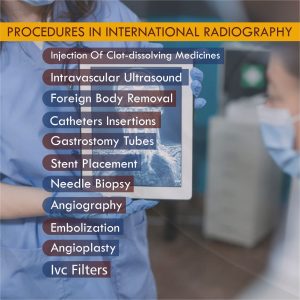
IVC filters: The doctor puts a small filter into the inferior vena cava (IVC). This is a large vein
in your abdomen. The filter catches blood clots that may go into your lungs
Injection of clot-dissolving medicines: The doctor injects clot-dissolving medicines such
as tissue plasminogen activator. This medicine dissolves blood clots and increases blood
flow to your arms, legs, or organs in your body.
Catheters insertions: The doctor puts a catheter into a large vein to give chemotherapy
medicines, nutrition, or hemodialysis. He or she may also put in a catheter before a
bone-marrow transplant.
Read more on Interventional Radiography.
RADIATION ONCOLOGY
This is otherwise known as Radiation Therapy. Radiation therapy is a cancer treatment that
uses high-energy x-ray or other particles to destroy cancer cells. A doctor who specializes in
giving radiation therapy to treat cancer is called a radiation oncologist.
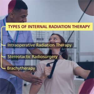
Three common types of internal radiation therapy include:
● Brachytherapy
● Intraoperative radiation therapy
● Stereotactic radiosurgery
Brachytherapy: It involves radioactive material that is implanted in the body. Dozens of tiny
“seeds” containing radioactive iodine are placed at the tumor site with a special needle or
catheter. This is done as an outpatient procedure. Brachytherapy is used for treatment of
prostate, cervical, endometrial, vaginal and breast cancers.
Intraoperative radiation therapy (IORT): It is used to treat an exposed tumor during cancer
surgery. IORT delivers a high dose of radiation to a surgically exposed treatment area.
Surrounding healthy organs and tissues are protected by lead shields. This type of radiation
can be used for certain gastrointestinal cancers and other cancers that are challenging to
remove during surgery.
Stereotactic radiosurgery (SRS): It is not actually surgery. Instead, it uses dozens of tiny
radiation beams to treat tumors in the head and neck with a single radiation dose. Gamma
Knife is used to treat cancer that has spread to the brain or head or neck area, as well as
tumors in the base of the skull, malignant gliomas, acoustic neuromas, pituitary tumors and
meningiomas.
Read more on Radiation Oncology
Here, professions in Radiography have been duly highlighted and elucidated to its simplest.
Making a choice of specialization, picking a career path should not be a stress now, right?